
 By
By 
This is an excerpt from The Curbside Guide, page 121, Adrenal Tumors
Description
An adrenal mass is suspected when the maximum width of the adrenal gland exceeds 1.5 cm, there is loss of normal architecture or shape, or the shape or size between the affected adrenal gland and the contralateral gland is asymmetrical. The latter comprise the initial criteria for diagnosis; however, a bulbous enlargement of the cranial or caudal pole of the adrenal gland is common in dogs with no adrenal pathology and can be misinterpreted as an adrenal mass.
If the suspected mass is not precipitating obvious signs (i.e., aggressive behavior), then an abdominal ultrasound should be repeated to confirm that the mass is a consistent finding before pursuing further diagnostics or surgery. Large breeds (Poodles, German Shepherds, Retrievers, and Terriers) and females appear to be overrepresented in the clinical reviews of adrenal tumors. Adrenal tumors in cats are rare with minimal information to characterize the disease. However, adrenal carcinoma and aldosterone producing tumors are the more common adrenal masses in our archived feline population. More specific information regarding this pathology may be found in the Feline Hyperaldosterone chapter.
Incidental adrenal lesions should be investigated clinically if discovered on ultrasound. Non-neoplastic adrenal lesions, such as cysts or granulomas, are very rare in dogs and cats, and the high incidence of metastatic lesions justifies a thorough hormonal screening as well as evaluation for non-adrenal neoplasms. Although incidental adrenal masses may appear to be nonfunctional at the time of diagnosis, it seems more likely that they are in fact subclinically functional. The diagnosis of functional adrenal tumors is discussed below; however, the identification of a nonfunctional, incidental adrenal mass creates a management dilemma.
Clinical Signs:
Clinical signs attributable to adrenal tumors are dependent on hormone secretion type. Please see below.
Diagnostics:
Cortical adrenal tumors, such as adenomas and adenocarcinomas, are responsible for 15–20% of hyperadrenocortical cases — what are commonly referred to as adrenal-dependent hyperadrenocortism (ADH) — in dogs.
The remaining tumors are the result of pituitary-dependent secretions, which give rise to pituitary-dependent hyperadrenocorticism (PDH). PDH cases tend to demonstrate bilateral hypertrophy with excessive adrenal length and, probably more importantly, width. These enlarged adrenal glands do not invade surrounding vascular structures and are defined by overstimulation resulting from excessive ACTH secretion from the pituitary gland. Yet, ADH cases are usually unilateral (bilateral in 10–20% of cases), may invade the aorta on the left or the vena cava on the right, and metastasize to the liver and lungs most frequently. Practitioners must differentiate ADH masses from hyperplastic, non-functional, benign adrenal tumors, as well as pheochromocytomas. Thus, dynamic function tests (ex. LDDS, HDDS, ACTH stimulation, ACTH baseline, urine cortisol-creatinine ratio) are essential, as is conducting routine biochemistry (ALP is elevated in more than 90% of cases) and urinalysis (true polyuria/polydipsia [PU/PD] with USG < 1.020) to determine adequately the need for surgical intervention or aggressive medical therapy. It is important to assess the following: blood pressure for hypertension; oscillating hyper- and hypotensive episodes in cases of pheochromocytomas; urine protein-creatinine ratios; and serum antithrombin III to determine the risk for thromboembolism. Moreover, it is essential to evaluate the entire clinical picture and objective probabilities of possessing a true hyperadrenocorticism case. This further entails ruling out other sources of PU/PD, such as primary polydipsia, renal disease, electrolyte abnormalities, infections, and diabetes insipidus or mellitus.
Malignant or Benign, Functional or Non-Functional: How to Decide?
In some cases, it may be difficult to determine whether the mass is malignant or benign, functional or nonfunctional, prior to surgical removal and histopathological examination. A thorough review of the clinical signs, physical examination findings, routine blood work, urine tests, and appropriate hormonal tests should be conducted to determine the functional status of an incidental adrenal mass.
Malignancy is more often associated with larger masses. The larger the mass, the more likely metastasis has already occurred, in spite of a lack of detectable lesions on ultrasound and thoracic radiographs. Invasion of the mass into surrounding organs or blood vessels also supports malignancy, as does the detection of additional mass lesions with abdominal ultrasound and thoracic radiographs. Use of imaging modalities, such as CT and MRI, will likely provide additional data on the characteristics of specific adrenal lesions for use in diagnosis and treatment planning.
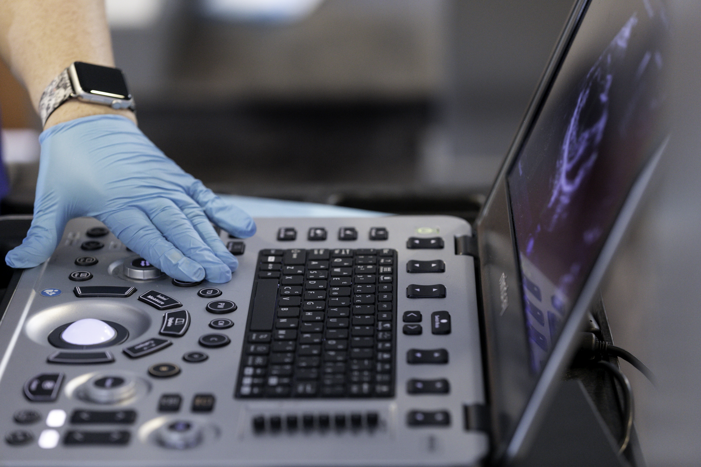
Ultrasonography is the primary instrument for assessing tumor size, aggressiveness, non-capsulated versus capsulated appearance, vascular invasion, and hepatic or other metastasis. Ideally, the patient will have fasted prior to the ultrasound; one may choose to administer an enema to enhance visibility around the ascending and descending colon. Ultrasound-guided biopsy or fine needle aspiration (FNA) may be possible on the larger masses, especially on the left side; however, adjacent vascular structures often prevent the feasibility of this procedure and is an operator dependent maneuver.


